Discover 8 critical signs of vitamin toxicity in reptiles. Learn prevention tips, treatment options, and expert veterinary advice to keep your scaly companions healthy.
Table of Contents
Reptile ownership has surged dramatically over the past decade, with over 4.5 million households in the United States keeping reptiles as pets according to the American Pet Products Association. While these fascinating creatures make wonderful companions, their unique nutritional needs often leave even experienced keepers confused about proper supplementation. Unfortunately, this confusion frequently leads to one of the most preventable yet serious health issues in captive reptiles: vitamin toxicity.
Vitamin toxicity in reptiles occurs when these animals receive excessive amounts of vitamins, particularly fat-soluble vitamins like A, D3, E, and K. Unlike mammals, reptiles have evolved specific metabolic pathways that make them particularly susceptible to vitamin overdoses. What makes this condition especially concerning is that the signs often develop gradually, making early detection challenging for even the most observant reptile keepers.
Understanding the warning signs of vitamin toxicity could literally save your reptile’s life. This comprehensive guide will explore the eight most critical symptoms every reptile owner must recognize, backed by veterinary expertise and real-world case studies. Whether you’re a beginner snake owner or an experienced iguana breeder, this information will help you provide the best possible care for your scaly companions.
Understanding Vitamin Toxicity in Reptiles: The Hidden Danger
Vitamin toxicity, also known as hypervitaminosis, represents one of the most misunderstood aspects of reptile husbandry. Unlike vitamin deficiencies, which have been extensively studied and documented, vitamin toxicity often flies under the radar until serious complications arise. Dr. Sarah Martinez, a board-certified exotic animal veterinarian with over 15 years of experience, explains: “I see more cases of vitamin toxicity than deficiency in my practice, primarily because well-meaning owners assume more vitamins equals better health.”
The physiology of reptiles makes them uniquely vulnerable to vitamin overdoses. Their slower metabolic rates mean vitamins accumulate in tissues over longer periods, and their liver’s limited capacity to process excess nutrients creates a perfect storm for toxicity. Fat-soluble vitamins (A, D3, E, and K) pose the greatest risk because they’re stored in fatty tissues and the liver rather than being excreted in urine like water-soluble vitamins.
Recent research published in the Journal of Exotic Pet Medicine found that nearly 30% of reptile health issues in captivity stem from nutritional imbalances, with vitamin toxicity accounting for approximately 40% of these cases. The study followed 500 reptiles across five years, revealing that bearded dragons, iguanas, and tortoises showed the highest susceptibility to vitamin A and D3 toxicity.
The most dangerous aspect of vitamin toxicity is its insidious nature. Unlike acute poisoning, which produces immediate symptoms, vitamin toxicity develops over weeks or months. This gradual onset means that by the time obvious symptoms appear, significant organ damage may have already occurred. Understanding this timeline is crucial for prevention and early intervention.
The Science Behind Reptile Vitamin Metabolism
To truly understand vitamin toxicity in reptiles, we must first examine how these animals process vitamins differently from mammals. Reptiles have evolved unique metabolic pathways that reflect their natural environments and dietary habits. In the wild, most reptiles consume whole prey items or varied plant matter, providing balanced nutrition without the risk of vitamin overdose.
Vitamin A toxicity occurs when reptiles receive excessive amounts of preformed vitamin A (retinol) rather than beta-carotene, which the body converts to vitamin A as needed. Many commercial reptile supplements contain high levels of preformed vitamin A, which can quickly accumulate to toxic levels. The liver stores vitamin A in stellate cells, and when these cells become overloaded, they release inflammatory compounds that damage liver tissue.
Vitamin D3 toxicity presents an even greater challenge because reptiles naturally produce vitamin D3 through UV exposure. When supplemental vitamin D3 is added to an already adequate UVB lighting setup, the combination can create dangerous calcium absorption rates. This leads to hypercalcemia, where excess calcium deposits in soft tissues, particularly the kidneys and cardiovascular system.
The metabolic rate variations among reptile species further complicate vitamin requirements. A bearded dragon basking at 95°F will metabolize vitamins much faster than a ball python maintained at 78°F. This difference means that standardized supplementation protocols often fail to account for species-specific metabolic needs.
Sign #1: Swollen or Puffy Appearance Around the Eyes and Head
One of the earliest and most recognizable signs of vitamin toxicity in reptiles is facial swelling, particularly around the eyes and temporal regions. This symptom most commonly indicates vitamin A toxicity, though it can also occur with vitamin D3 overdose. The swelling results from fluid accumulation in tissues due to compromised liver function and altered protein metabolism.
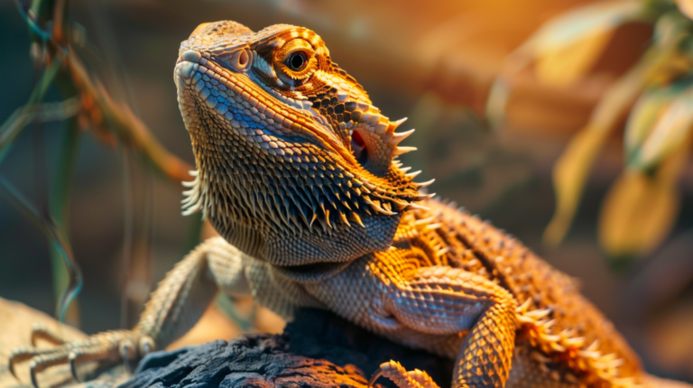
In bearded dragons, vitamin A toxicity typically manifests as bilateral periorbital swelling, giving the animal a “bug-eyed” appearance. The swelling may be subtle initially, appearing as slightly puffy eyelids that gradually worsen over several weeks. Owners often mistake this for minor irritation or shedding-related issues, delaying necessary veterinary intervention.
Iguanas with vitamin A toxicity may develop more generalized facial swelling, including enlarged temporal muscles and swollen jowls. This can significantly impact their ability to eat and drink normally. The swelling often feels firm to the touch and may cause the animal to hold its head at unusual angles to compensate for impaired vision.
Tortoises and turtles present unique challenges because their natural head shape can mask early swelling. However, careful observation reveals asymmetrical swelling around the eyes, difficulty retracting the head fully into the shell, and reluctance to extend the head for feeding. Box turtles with vitamin A toxicity may show unilateral swelling initially, which can progress to bilateral involvement if left untreated.
The severity of facial swelling correlates directly with the degree of vitamin toxicity and duration of exposure. Mild cases may resolve with dietary modifications and supportive care, while severe cases require aggressive treatment including diuretics, anti-inflammatory medications, and intensive monitoring. Recovery time varies significantly, with some animals showing improvement within days while others may take weeks to return to normal appearance.
Sign #2: Changes in Skin Color and Texture
Vitamin toxicity profoundly affects reptile skin health, producing distinctive changes in coloration and texture that experienced keepers learn to recognize quickly. These integumentary changes occur because vitamins play crucial roles in cellular metabolism and collagen synthesis, and excess amounts disrupt normal skin renewal processes.
Vitamin A toxicity often causes skin to become unnaturally pale or develop a yellowish tint, particularly noticeable in normally vibrant species like bearded dragons or leopard geckos. The skin may appear thin and translucent, with visible blood vessels beneath the surface. In severe cases, the skin becomes fragile and prone to tearing with minimal handling.
Hyperkeratosis, or thickening of the outer skin layer, represents another common manifestation of vitamin toxicity. This condition causes the skin to develop a rough, sandpaper-like texture and may lead to abnormal shedding patterns. Snakes with vitamin A toxicity may experience stuck sheds, particularly around the eyes and tail tip, while lizards may develop thick, flaky patches that resist normal shedding.
Color changes in reptiles with vitamin toxicity often follow predictable patterns. Green iguanas may lose their vibrant emerald coloration, becoming dull olive or brown. Bearded dragons might develop muddy, washed-out colors instead of their typical bright yellows and oranges. These changes occur because excess vitamins interfere with chromatophore function, the specialized cells responsible for color production.
The texture changes associated with vitamin toxicity extend beyond surface appearance. Affected skin often feels unusually dry or, conversely, may develop an oily consistency. Some reptiles develop small, hard nodules under the skin, particularly around the neck and limbs. These nodules represent areas where excess vitamins have crystallized in tissue.
Monitoring skin changes requires regular, close observation of your reptile. Weekly photography can help document subtle changes that might otherwise go unnoticed. Pay particular attention to areas that typically show the most vibrant coloration, as these regions often display the earliest signs of vitamin-related skin problems.
Sign #3: Lethargy and Reduced Activity Levels
Behavioral changes, particularly decreased activity and lethargy, represent some of the most concerning signs of vitamin toxicity in reptiles. Unlike mammals, reptiles cannot verbally communicate discomfort, making behavioral observation crucial for early detection of health problems. Vitamin toxicity affects cellular energy production and neurological function, leading to pronounced changes in normal behavior patterns.
The onset of lethargy in vitamin-toxic reptiles typically follows a predictable progression. Initially, owners may notice their pet spending slightly more time hiding or basking than usual. This subtle change often gets dismissed as normal variation in behavior. However, as toxicity progresses, the animal becomes increasingly inactive, showing reluctance to explore, hunt, or engage in normal territorial behaviors.
Bearded dragons with vitamin toxicity may abandon their characteristic head-bobbing and arm-waving displays, instead remaining motionless for extended periods. They may fail to achieve their normal basking positions or seem unable to maintain proper posture while basking. The bright, alert demeanor typical of healthy bearded dragons gives way to a dull, listless appearance.
Ball pythons and other snake species present particular challenges because their naturally sedentary nature can mask early signs of lethargy. However, careful observation reveals subtle differences in movement quality, response to handling, and feeding behavior. Toxic snakes may remain in the same position for days without the normal repositioning that healthy snakes display.
Aquatic species like red-eared sliders show lethargy through reduced swimming activity and prolonged basking periods. They may float motionlessly at the surface instead of actively swimming and diving. Terrestrial turtles may stop exploring their enclosures and remain withdrawn in their shells for extended periods.
The neurological effects of vitamin toxicity contribute significantly to behavioral changes. Excess vitamin A can cause cerebral edema, leading to confusion, disorientation, and altered consciousness. Animals may appear “drunk” or uncoordinated, with delayed responses to stimuli. Some reptiles develop abnormal sleep patterns, sleeping during normally active periods or showing restlessness during typical rest times.
Sign #4: Loss of Appetite and Feeding Difficulties
Appetite changes represent one of the most reliable indicators of vitamin toxicity in reptiles, though the specific presentation varies considerably among species and individuals. The gastrointestinal system bears the brunt of vitamin toxicity effects, with liver damage, altered digestive enzyme production, and gastric irritation all contributing to feeding problems.
The progression of appetite loss typically follows a predictable pattern. Initially, reptiles may show decreased enthusiasm for food, taking longer to respond to feeding cues or requiring more encouragement to eat. This subtle change often coincides with the early stages of vitamin accumulation in tissues. As toxicity progresses, animals may refuse preferred foods while still accepting highly palatable items, eventually progressing to complete anorexia.
Bearded dragons with vitamin A toxicity often develop a characteristic feeding pattern where they approach food items but fail to consume them. They may show normal hunting behaviors, stalking and positioning for strikes, but lack the coordination or motivation to complete feeding. This behavior results from the neurological effects of vitamin toxicity combined with oral discomfort from tissue swelling.
Snake species present unique challenges because their feeding frequency naturally varies with temperature, season, and individual factors. However, vitamin-toxic snakes typically show complete food refusal even when offered appropriately sized prey items. The normal feeding response, including tongue flicking and exploratory behavior, becomes diminished or absent entirely.
Herbivorous reptiles like iguanas and tortoises may show selective appetite loss, refusing certain food items while accepting others. This selectivity often reflects altered taste perception due to vitamin toxicity. Some animals develop pica, consuming inappropriate items like substrate or non-food objects, possibly in response to nutritional imbalances caused by impaired vitamin metabolism.
The physical mechanics of feeding may also become compromised in vitamin-toxic reptiles. Oral swelling from vitamin A toxicity can make it difficult or impossible to open the mouth adequately for feeding. Some animals may attempt to eat but drop food items due to weakened jaw muscles or altered oral sensation.
Sign #5: Abnormal Shedding Patterns
Shedding abnormalities represent one of the most visible and diagnostically significant signs of vitamin toxicity in reptiles. Normal shedding depends on precise hormonal regulation, adequate hydration, and healthy cellular turnover—all processes that vitamin toxicity disrupts. Understanding normal shedding patterns for your specific species is crucial for recognizing when problems arise.
Vitamin A toxicity particularly affects shedding because this vitamin plays essential roles in epithelial cell development and differentiation. Excess vitamin A accelerates cellular turnover, leading to more frequent shedding cycles. However, the quality of sheds decreases dramatically, with incomplete or patchy shedding becoming the norm rather than the exception.
Snakes with vitamin toxicity may develop dysecdysis, where the old skin fails to shed properly and remains attached in patches. The retained skin often appears dry, tight, and discolored, particularly around the eyes (spectacle retention) and tail tip. These areas require high cellular turnover rates and are therefore most susceptible to vitamin-related disruption.
Lizards may experience similar shedding difficulties, but their continuous shedding process makes problems less immediately obvious. Look for areas where old skin remains attached for extended periods, particularly around the toes, tail, and limb joints. These retained skin patches can constrict blood flow and lead to tissue necrosis if not addressed promptly.
The timing of shedding cycles also becomes disrupted with vitamin toxicity. Young reptiles that normally shed every few weeks may attempt to shed weekly or even more frequently. Conversely, some animals may go extended periods without shedding, accumulating multiple layers of old skin that create a dull, unhealthy appearance.
Shedding quality provides important diagnostic information. Healthy sheds come off in large, intact pieces (complete body tubes in snakes), while vitamin-toxic animals produce fragmented, brittle shed pieces. The shed skin itself may appear unusually thin or thick, with abnormal coloration or texture that reflects the underlying metabolic disruption.
Sign #6: Respiratory Distress and Breathing Difficulties
Respiratory symptoms in reptiles with vitamin toxicity often develop as secondary complications but can quickly become life-threatening if not addressed promptly. The respiratory system becomes compromised through multiple mechanisms, including direct tissue damage from vitamin accumulation, secondary bacterial infections, and cardiovascular complications that affect oxygen delivery.
Vitamin A toxicity particularly affects respiratory tissues because the vitamin plays crucial roles in maintaining mucous membrane integrity. Excess vitamin A causes hyperplasia of respiratory epithelium, leading to increased mucus production and narrowed airways. This creates ideal conditions for bacterial overgrowth and secondary respiratory infections.
Early respiratory signs include increased breathing rate and effort, particularly noticeable during periods of activity or stress. Reptiles may adopt unusual postures to facilitate breathing, such as extending the neck, opening the mouth, or positioning themselves to maximize airflow. These compensatory behaviors indicate significant respiratory compromise.
Audible breathing sounds represent more advanced respiratory distress. Wheezing, clicking, or rattling sounds during inhalation or exhalation suggest significant airway obstruction or fluid accumulation. These sounds may be subtle initially but typically worsen over time without intervention.
Mouth breathing in reptiles normally indicates severe respiratory distress because these animals prefer nasal breathing when possible. Vitamin-toxic reptiles may keep their mouths slightly open or display obvious respiratory effort with each breath. This behavior becomes particularly pronounced during periods of increased oxygen demand.
The cardiovascular effects of vitamin D3 toxicity can further compromise respiratory function. Hypercalcemia from excessive vitamin D3 can cause arrhythmias and decreased cardiac output, reducing the efficiency of oxygen delivery to tissues. This creates a cascade effect where respiratory symptoms worsen despite adequate lung function.
Sign #7: Digestive Issues and Unusual Bowel Movements
Gastrointestinal symptoms frequently accompany vitamin toxicity in reptiles, reflecting the complex relationship between vitamin metabolism and digestive function. The liver, which processes and stores many vitamins, becomes overwhelmed in cases of toxicity, leading to impaired bile production and altered digestive enzyme function.
Diarrhea represents one of the most common digestive symptoms in vitamin-toxic reptiles. The consistency, color, and frequency of bowel movements provide valuable diagnostic information. Vitamin A toxicity often produces yellowish, greasy stools due to impaired fat digestion, while vitamin D3 toxicity may cause watery diarrhea with unusual coloration.
The timing of digestive symptoms relative to feeding provides important clues about the underlying problem. Reptiles with vitamin toxicity may experience delayed gastric emptying, leading to regurgitation or vomiting hours or days after feeding. This delayed response occurs because impaired liver function affects the production of digestive enzymes and bile acids necessary for proper food breakdown.
Constipation may also occur in vitamin-toxic reptiles, particularly in species that normally produce regular, well-formed stools. The altered intestinal motility and changed bacterial populations in the gut can lead to prolonged transit times and impacted fecal material. This is especially problematic in tortoises and large lizards that rely on regular elimination for optimal health.
The appearance of blood in stool indicates significant gastrointestinal irritation and potential tissue damage. Vitamin toxicity can cause direct irritation of intestinal mucosa, leading to inflammation and bleeding. This symptom requires immediate veterinary attention as it may indicate severe internal damage.
Changes in urate production also accompany vitamin toxicity. Reptiles typically produce white, chalky urates along with their feces, but vitamin-toxic animals may produce unusually colored urates (yellow, orange, or green) or may show increased or decreased urate volume. These changes reflect altered kidney function and protein metabolism.
Sign #8: Neurological Symptoms and Behavioral Changes
Neurological symptoms represent some of the most serious manifestations of vitamin toxicity in reptiles, often indicating advanced systemic involvement that requires immediate veterinary intervention. The nervous system’s high metabolic demands and complex vitamin requirements make it particularly vulnerable to both deficiency and toxicity states.
Vitamin A toxicity can cause increased intracranial pressure due to cerebral edema, leading to characteristic neurological signs. Affected reptiles may show head tilting, circling behaviors, or difficulty maintaining normal orientation. These symptoms occur because excess vitamin A disrupts normal brain cell function and increases fluid accumulation within the skull.
Seizure activity, while less common, represents a severe neurological manifestation of vitamin toxicity. Reptiles may experience tonic-clonic seizures, focal seizures affecting specific body regions, or subtle absence seizures that appear as brief periods of unresponsiveness. Any seizure activity in a reptile requires immediate emergency veterinary care.
Coordination problems become evident as toxicity progresses. Affected animals may show ataxia (unsteady gait), difficulty climbing or maintaining balance, or unusual positioning of limbs. Snakes may develop abnormal undulation patterns or difficulty maintaining proper body posture during movement.
Altered consciousness represents a grave neurological sign in vitamin-toxic reptiles. Animals may appear dull, unresponsive, or show delayed reactions to stimuli. Some reptiles develop stupor or coma-like states, indicating severe brain dysfunction that requires aggressive treatment.
Visual disturbances commonly accompany vitamin A toxicity due to retinal damage and increased intraocular pressure. Affected reptiles may show reluctance to hunt, difficulty targeting food items, or abnormal pupil responses to light. These visual changes may be reversible with early treatment but can become permanent if toxicity persists.
Temperature regulation problems may also develop as neurological toxicity progresses. The hypothalamus, which controls thermoregulation, becomes affected by vitamin accumulation, leading to inappropriate behavioral thermoregulation. Reptiles may choose inappropriate temperatures or show little interest in thermoregulatory behavior.
Prevention Strategies: Protecting Your Reptile from Vitamin Toxicity
Prevention remains the most effective approach to managing vitamin toxicity in reptiles. Understanding proper supplementation protocols, recognizing species-specific requirements, and maintaining appropriate environmental conditions significantly reduce the risk of vitamin-related health problems. The key lies in achieving nutritional balance rather than maximizing vitamin intake.
Species-specific research forms the foundation of effective prevention. Each reptile species has evolved unique nutritional requirements based on their natural diet, habitat, and metabolic characteristics. Bearded dragons, for example, require different vitamin A levels than ball pythons due to their omnivorous versus carnivorous diets. Consulting species-specific care guides and working with experienced reptile veterinarians ensures appropriate supplementation protocols.
UVB lighting plays a crucial role in preventing vitamin D3 toxicity while maintaining adequate vitamin D metabolism. Proper UVB exposure allows reptiles to synthesize vitamin D3 naturally, reducing or eliminating the need for supplemental vitamin D3. However, combining supplemental vitamin D3 with UVB lighting can quickly lead to toxicity. Monitor UVB bulb output regularly and replace bulbs according to manufacturer recommendations.
Dietary diversity prevents nutritional imbalances that can contribute to vitamin toxicity. Wild reptiles consume varied diets that provide balanced nutrition without excessive vitamin concentrations. Captive reptiles benefit from similarly diverse diets that include appropriate prey items, vegetables, and commercial foods. Avoid over-reliance on vitamin-fortified commercial diets, which can contribute to vitamin accumulation.
Quality control in supplement selection significantly impacts toxicity risk. Choose supplements from reputable manufacturers that provide detailed ingredient lists and vitamin concentrations. Avoid products with excessive vitamin A or D3 levels, and be particularly cautious with supplements marketed as “high-potency” or “super-concentrated.” Many reptile health problems stem from well-intentioned but excessive supplementation.
Treatment Approaches: What to Do When Toxicity Occurs
When vitamin toxicity is suspected, immediate veterinary intervention provides the best chance for successful treatment and recovery. The treatment approach depends on the specific vitamins involved, the severity of symptoms, and the species affected. Early intervention dramatically improves outcomes, while delayed treatment may result in permanent organ damage or death.
Veterinary diagnosis typically involves comprehensive blood work to assess vitamin levels, liver function, and kidney function. Vitamin A levels can be measured directly, while vitamin D3 toxicity is diagnosed through elevated blood calcium and phosphorus levels. Additional diagnostic tests may include radiographs to assess bone density and organ involvement.
Treatment protocols focus on stopping vitamin intake, supporting organ function, and managing symptoms. The first step involves immediately discontinuing all vitamin supplementation and reviewing the diet to eliminate vitamin-fortified foods. This intervention alone may be sufficient for mild cases, but more severe toxicity requires aggressive medical management.
Supportive care forms the backbone of toxicity treatment. Fluid therapy helps maintain hydration and supports kidney function in eliminating excess vitamins. Anti-inflammatory medications may be necessary to reduce tissue swelling and inflammation. Some cases require hospitalization for intensive monitoring and treatment.
Specific antidotes exist for certain types of vitamin toxicity. Vitamin A toxicity may respond to retinoid-binding therapy, while vitamin D3 toxicity requires medications to reduce calcium absorption and increase calcium excretion. These treatments must be carefully monitored to avoid creating secondary nutritional deficiencies.
Recovery time varies significantly depending on the severity of toxicity and the reptile’s overall health status. Mild cases may show improvement within days to weeks, while severe toxicity can require months of treatment and monitoring. Some effects, particularly neurological damage, may be permanent despite appropriate treatment.
Working with Veterinarians: Building a Healthcare Team
Establishing a relationship with a qualified exotic animal veterinarian before health problems arise provides the best foundation for successful reptile care. Not all veterinarians have experience with reptiles, so finding a practitioner with specific exotic animal training and experience is crucial for optimal care.
Board certification in exotic animal medicine represents the highest level of specialized training available. Veterinarians with this certification have completed additional residency training and passed rigorous examinations in exotic animal medicine. While not always available in all areas, board-certified exotic veterinarians provide the most advanced level of care for reptiles.
Regular wellness examinations allow veterinarians to establish baseline health parameters and identify problems before they become serious. Annual examinations for adult reptiles and semi-annual examinations for young, elderly, or chronically ill animals provide optimal preventive care. These visits should include nutritional counseling and husbandry review.
Emergency preparedness includes knowing how to contact your veterinarian outside of normal business hours and having a plan for emergency transportation. Some cities have 24-hour emergency veterinary clinics with exotic animal capabilities, while others may require travel to university veterinary hospitals or specialized practices.
Building a relationship with your veterinarian involves open communication about your reptile’s care, any concerns you may have, and changes in the animal’s behavior or appearance. Don’t hesitate to ask questions about nutrition, supplementation, or husbandry practices. A good veterinarian welcomes these discussions and uses them to provide better care for your pet.
Species-Specific Considerations for Common Pet Reptiles
Different reptile species exhibit unique susceptibilities to vitamin toxicity based on their evolutionary adaptations and natural dietary preferences. Understanding these species-specific factors helps reptile keepers tailor their supplementation and husbandry practices to minimize toxicity risks while maintaining optimal health.
Bearded Dragons show particular susceptibility to vitamin A toxicity due to their omnivorous diet and high growth rates. Young bearded dragons require more frequent feeding and may receive excessive vitamin supplementation during their rapid growth phase. The combination of vitamin-dusted insects, vitamin-fortified commercial diets, and additional supplements can quickly lead to toxicity. Limiting vitamin A supplementation to 2-3 times per week and focusing on beta-carotene-rich vegetables helps prevent toxicity.
Ball Pythons and Other Snakes primarily face vitamin D3 toxicity risks when UVB lighting is combined with vitamin D3 supplementation. Since snakes typically consume whole prey items that provide balanced nutrition, additional vitamin supplementation is rarely necessary. However, some keepers supplement prey items with vitamin powders, creating toxicity risks. Frozen-thawed prey items maintain adequate vitamin content without requiring additional supplementation.
Leopard Geckos demonstrate intermediate susceptibility to vitamin toxicity, with most problems arising from excessive vitamin D3 supplementation. These nocturnal species don’t require UVB lighting, making them dependent on dietary vitamin D3. However, over-supplementation combined with calcium-rich diets can quickly lead to hypercalcemia. Limiting vitamin D3 supplementation to once weekly typically provides adequate nutrition without toxicity risks.
Green Iguanas face unique challenges due to their herbivorous diet and large size. These reptiles require significant amounts of vitamin A from plant sources, but supplemental vitamin A can quickly accumulate to toxic levels. Focus on providing vitamin A through beta-carotene-rich vegetables like squash, sweet potato, and dark leafy greens rather than supplemental vitamin A.
Red-Eared Sliders and Aquatic Turtles often receive excessive vitamin supplementation due to concerns about nutritional deficiencies in commercial turtle pellets. However, high-quality commercial diets typically provide adequate vitamin levels without requiring additional supplementation. The combination of vitamin-fortified pellets, vitamin-dusted prey items, and liquid vitamin supplements frequently leads to toxicity.
Environmental Factors Affecting Vitamin Metabolism
Environmental conditions significantly influence vitamin metabolism in reptiles, affecting both the risk of toxicity and the effectiveness of treatment. Temperature, humidity, lighting, and seasonal variations all play crucial roles in how reptiles process and utilize vitamins. Understanding these environmental factors helps optimize husbandry practices to minimize toxicity risks.
Temperature directly affects metabolic rate and vitamin utilization in reptiles. Higher temperatures increase metabolic rate, leading to faster vitamin processing and potentially higher vitamin requirements. However, this increased metabolism also means that toxic levels can be reached more quickly. Maintaining appropriate temperature gradients allows reptiles to thermoregulate effectively and optimize their vitamin metabolism.
UVB Lighting represents one of the most critical environmental factors affecting vitamin D metabolism. Proper UVB exposure allows reptiles to synthesize vitamin D3 naturally while preventing the accumulation of excessive supplemental vitamin D3. However, inadequate UVB combined with vitamin D3 supplementation can lead to deficiency, while excessive UVB combined with supplementation can cause toxicity.
Seasonal Variations affect vitamin requirements in many reptile species. Some species naturally experience seasonal changes in appetite, activity, and vitamin requirements. Continuing the same supplementation schedule year-round may lead to toxicity during periods of reduced activity and vitamin utilization.
Humidity affects skin health and shedding patterns, which can influence vitamin A requirements. Low humidity can increase vitamin A needs for maintaining healthy skin, while excessive humidity combined with high vitamin A supplementation may contribute to skin problems.
Stress from inappropriate environmental conditions can affect vitamin metabolism and increase susceptibility to toxicity. Chronic stress elevates cortisol levels, which can interfere with vitamin absorption and utilization. Maintaining appropriate environmental conditions reduces stress and supports optimal vitamin metabolism.
Long-Term Management and Monitoring
Successful long-term management of reptiles with a history of vitamin toxicity requires ongoing vigilance and systematic monitoring. Recovery from vitamin toxicity can be prolonged, and some animals may have increased susceptibility to future episodes. Developing a comprehensive monitoring plan helps ensure continued health and prevents recurrence.
Regular Health Assessments should include weekly evaluations of appetite, activity level, and general appearance. Document any changes in behavior, feeding response, or physical appearance to identify potential problems early. Monthly weight monitoring helps track overall health trends and identify gradual changes that might indicate developing problems.
Photographic Documentation provides valuable objective records of your reptile’s condition over time. Weekly photographs taken under consistent lighting conditions can reveal subtle changes in skin color, texture, or body condition that might otherwise be missed. These photos also provide valuable information for veterinary consultations.
Feeding Records help identify patterns in appetite and feeding behavior that might indicate developing health problems. Record the types and amounts of food offered, food items consumed, and any supplements provided. This information proves invaluable for veterinary diagnosis and treatment planning.
Environmental Monitoring ensures consistent conditions that support optimal health. Track temperature, humidity, and UVB output regularly to identify any environmental factors that might contribute to health problems. Replace UVB bulbs according to manufacturer recommendations and monitor their output with appropriate meters.
Veterinary Follow-Up schedules should be established based on the severity of previous toxicity episodes and the animal’s current health status. Animals with a history of severe toxicity may require more frequent examinations and blood work to monitor organ function and vitamin levels.
Frequently Asked Questions
Q: How quickly can vitamin toxicity develop in reptiles?
A: Vitamin toxicity can develop over different timeframes depending on the vitamin involved and the degree of over-supplementation. Fat-soluble vitamins like A and D3 can accumulate over weeks to months, while water-soluble vitamins are generally excreted more quickly. Acute toxicity from massive overdoses can occur within days, but chronic toxicity from regular over-supplementation typically develops over 4-12 weeks.
Q: Can vitamin toxicity be completely reversed with treatment?
A: The reversibility of vitamin toxicity depends on the severity and duration of exposure. Mild cases caught early often resolve completely with appropriate treatment. However, severe toxicity that has caused organ damage, particularly to the liver or kidneys, may result in permanent health problems. Neurological damage from vitamin A toxicity can be particularly persistent, though some improvement may occur over months of treatment.
Q: Is it safer to under-supplement rather than over-supplement vitamins?
A: While both vitamin deficiency and toxicity can cause serious health problems, mild under-supplementation is generally safer than over-supplementation for most reptiles. Many reptiles can obtain adequate nutrition from a well-balanced diet with minimal supplementation. However, certain species and life stages (such as growing juveniles or gravid females) may have higher vitamin requirements that necessitate careful supplementation.
Q: How do I know if my reptile’s commercial diet already contains adequate vitamins?
A: Check the ingredient list and guaranteed analysis on commercial reptile foods to determine vitamin content. High-quality commercial diets typically provide adequate vitamin levels for most species. If the diet contains significant amounts of vitamin A or D3, additional supplementation may not be necessary. Consult with an exotic animal veterinarian to determine whether your reptile’s diet requires additional supplementation.
Q: What should I do if I suspect I’ve been over-supplementing my reptile?
A: If you suspect over-supplementation, immediately discontinue all vitamin supplements and schedule a veterinary examination. Document any symptoms you’ve observed and bring information about the supplements you’ve been using, including brand names, dosages, and frequency of administration. Early veterinary intervention provides the best chance for successful treatment and recovery.
Q: Are there any reptile species that are particularly resistant to vitamin toxicity?
A: While no reptile species is completely immune to vitamin toxicity, some species appear more tolerant than others. Large, slow-growing species like adult tortoises may be somewhat more tolerant due to their slower metabolic rates. However, all reptiles can develop vitamin toxicity with sufficient exposure, so appropriate caution should be exercised regardless of species.
Expert Resources and Further Reading
For reptile keepers seeking additional information about vitamin toxicity and reptile nutrition, several authoritative resources provide evidence-based guidance. The Association of Reptilian and Amphibian Veterinarians (ARAV) offers comprehensive care guidelines and can help locate qualified exotic animal veterinarians in your area.
The Journal of Exotic Pet Medicine publishes cutting-edge research on reptile nutrition and health, including recent studies on vitamin toxicity. While some articles require technical knowledge to interpret, they provide the most current understanding of reptile nutritional needs and toxicity risks.
University extension programs often provide excellent resources for reptile husbandry and nutrition. Many veterinary schools with exotic animal programs offer continuing education courses for reptile keepers, covering topics like proper supplementation and recognizing health problems.
For more expert pet care tips and product recommendations, visit BlithePet.com — your trusted source for pet wellness.
Conclusion
Vitamin toxicity in reptiles represents a serious but preventable health threat that every reptile keeper must understand. The eight critical signs outlined in this guide—facial swelling, skin changes, lethargy, appetite loss, shedding problems, respiratory distress, digestive issues, and neurological symptoms—provide early warning signals that can save your reptile’s life when recognized promptly.
The key to preventing vitamin toxicity lies in understanding that more is not always better when it comes to reptile nutrition. Balanced supplementation based on species-specific requirements, combined with appropriate environmental conditions and regular veterinary care, provides the foundation for optimal reptile health. Remember that vitamin toxicity often develops gradually, making regular observation and monitoring essential for early detection.
Working with qualified exotic animal veterinarians, maintaining detailed health records, and staying informed about current reptile care practices all contribute to successful long-term reptile care. The investment in proper nutrition and preventive care pays dividends in the form of healthier, longer-lived reptiles that can provide years of companionship and enjoyment.
If you notice any of the warning signs discussed in this article, don’t delay in seeking veterinary attention. Early intervention dramatically improves treatment outcomes and reduces the risk of permanent organ damage. Remember that reptiles are masters at hiding illness, so subtle changes in behavior or appearance may represent significant underlying health problems.
The reptile-keeping community continues to evolve in its understanding of proper nutrition and supplementation. Stay connected with reputable reptile organizations, attend educational seminars, and maintain open communication with your veterinarian to ensure you’re providing the best possible care for your scaly companions. Your vigilance and dedication to proper husbandry practices make the difference between a reptile that merely survives in captivity and one that truly thrives.
Have a similar experience with your pet? Share it in the comments below!