Recognize respiratory infections in rodents early with these 7 critical breathing signs. Expert veterinary guide for hamsters, rats, mice, and guinea pigs.
Table of Contents
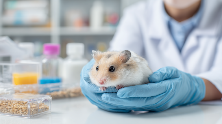
Every small pet owner’s worst nightmare is watching their beloved rodent struggle to breathe. Respiratory infections in rodents are among the most common yet potentially fatal health issues affecting hamsters, rats, mice, guinea pigs, and other small pets. Unlike larger animals, rodents have incredibly delicate respiratory systems that can deteriorate rapidly when infection strikes.
According to veterinary statistics, respiratory infections account for approximately 40% of all illness-related visits to exotic pet veterinarians, making them the leading cause of morbidity in captive rodent populations. The challenge lies in recognizing the subtle early warning signs before the infection progresses to life-threatening pneumonia or sepsis.
This comprehensive guide will help you identify the seven most serious breathing signs that indicate your rodent may be suffering from a respiratory infection, understand the underlying causes, and know when immediate veterinary intervention is necessary. Early detection and prompt treatment can mean the difference between a full recovery and tragic loss.
Understanding Rodent Respiratory Anatomy and Vulnerability
Before diving into the warning signs, it’s crucial to understand why rodents are particularly susceptible to respiratory infections. Their unique anatomical features create both advantages and vulnerabilities in their respiratory health.
The Delicate Respiratory System
Rodents possess remarkably efficient respiratory systems designed for their high metabolic rates. Their lungs are proportionally smaller than those of larger mammals, but they compensate with faster breathing rates and higher oxygen extraction efficiency. However, this efficiency comes at a cost – their respiratory passages are narrow and easily blocked by inflammation or secretions.
The nasal passages in rodents are intricate networks of turbinates that warm and humidify incoming air. While this system works beautifully under normal conditions, it becomes problematic when swelling occurs. Even minor inflammation can significantly restrict airflow, leading to mouth breathing and increased stress on the entire respiratory system.
Common Pathogens and Risk Factors
Respiratory infections in rodents are typically caused by bacterial pathogens such as Mycoplasma, Pasteurella, Bordetella, and Streptococcus species. Viral infections, while less common, can also trigger secondary bacterial infections that prove more challenging to treat.
Environmental factors play a significant role in respiratory infection development. Poor ventilation, high ammonia levels from inadequate cage cleaning, dusty bedding materials, and sudden temperature changes all contribute to increased infection risk. Stress from overcrowding, poor nutrition, or inadequate housing can further compromise the immune system, making rodents more susceptible to respiratory pathogens.
The 7 Serious Breathing Signs You Cannot Ignore
| # | Breathing Sign | Description & What to Look For | Severity | Action |
|---|---|---|---|---|
| 1 | Rapid, Shallow Breathing (Tachypnea) |
Breathing rate significantly faster than normal for the species. Visible rapid chest movements, often accompanied by restlessness and inability to settle comfortably. | Critical | Immediate vet care |
| 2 | Open-Mouth Breathing |
Rodent breathing through the mouth instead of nose. This is extremely abnormal as healthy rodents are obligate nose breathers. Often indicates severe respiratory distress. | Critical | Emergency vet visit |
| 3 | Audible Breathing Sounds |
Wheezing, clicking, crackling, or squeaking sounds during breathing. May be heard from across the room. Indicates airway obstruction or fluid in lungs. | Urgent | Same-day vet care |
| 4 | Abnormal Body Positioning |
Sitting hunched with head extended, refusing to lie down, or positioning to maximize airflow. Often accompanied by visible effort to breathe. | Urgent | Same-day vet care |
| 5 | Nasal Discharge & Facial Swelling |
Clear, colored, or bloody discharge from nose. May include swelling around eyes or face, crusting around nostrils, or frequent pawing at face. | Urgent | Within 24 hours |
| 6 | Reduced Activity & Lethargy |
Significant decrease in normal activities, reluctance to move, decreased appetite, and spending more time sleeping or hiding than usual. | Moderate | Monitor & vet consult |
| 7 | Changes in Vocalization |
Hoarse, raspy, or absent vocalizations. Some rodents may become unusually quiet, while others may make distressed sounds when breathing. | Moderate | Schedule vet visit |
1. Rapid, Shallow Breathing (Tachypnea)
The first and often most subtle sign of respiratory distress in rodents is an increase in breathing rate accompanied by shallow respirations. Normal breathing rates vary by species – rats typically breathe 70-110 times per minute, while hamsters may breathe 35-135 times per minute. However, when respiratory infection develops, you’ll notice the breathing becoming noticeably faster and more shallow.
This compensatory mechanism occurs as the body attempts to maintain adequate oxygen levels despite reduced lung capacity. The breathing pattern changes from the normal deep, rhythmic respirations to quick, inefficient gasps. You might observe your rodent’s chest rising and falling rapidly, even during rest periods.
Dr. Sarah Mitchell, a board-certified exotic veterinarian with over 15 years of experience, explains: “Tachypnea is often the first sign owners notice, but it’s frequently dismissed as normal activity. The key is observing your pet during quiet moments – if they’re breathing rapidly while resting, this warrants immediate attention.”
2. Open-Mouth Breathing
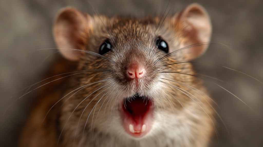
Perhaps the most alarming sign is when your rodent begins breathing through their mouth. Unlike cats and dogs, rodents are obligate nose breathers under normal circumstances. Open-mouth breathing indicates severe respiratory distress and suggests that nasal passages are significantly obstructed.
This breathing pattern is particularly dangerous because it bypasses the natural filtering, warming, and humidifying functions of the nasal passages. The mouth breathing creates a cycle of increased irritation and dehydration of the respiratory tract, potentially worsening the infection.
When you observe open-mouth breathing, you may also notice the tongue protruding slightly, excessive drooling, or a blue-tinged discoloration around the mouth and nose (cyanosis). These are emergency signs requiring immediate veterinary intervention.
3. Audible Breathing Sounds
Healthy rodents breathe silently, so any audible breathing sounds should raise immediate concern. These sounds can manifest as wheezing, crackling, squeaking, or clicking noises that become apparent when you listen closely to your pet or when they’re near your ear.
Wheezing typically indicates narrowed airways due to inflammation or mucus accumulation. The sound resembles a high-pitched whistle and is most noticeable during expiration. Crackling sounds suggest fluid accumulation in the lungs (pulmonary edema) and are often accompanied by other severe symptoms. Clicking or squeaking noises may indicate upper respiratory tract involvement, particularly nasal or throat infections.
These audible signs often worsen during activity or stress and may be accompanied by visible chest movement abnormalities. Recording these sounds on your phone can be helpful for your veterinarian’s assessment.
4. Abnormal Body Positioning
Rodents with respiratory infections often adopt specific postures to facilitate breathing. The most common position is sitting upright with the head extended forward and slightly elevated – a posture veterinarians call “orthopnea.” This position helps maximize chest expansion and reduces the work of breathing.
You might also observe your rodent avoiding lying down or frequently changing positions as if unable to get comfortable. Some animals will brace themselves against cage walls or accessories to assist with breathing. These behavioral changes indicate significant respiratory compromise and the body’s attempt to optimize airflow.
The “tripod position” is another concerning posture where the rodent sits with front legs extended forward, supporting their upper body weight. This position indicates severe respiratory distress and requires emergency veterinary care.
5. Nasal Discharge and Facial Swelling
Nasal discharge in rodents can range from clear and watery to thick, yellow, or green secretions. While minimal clear discharge might be normal in some circumstances, any persistent or colored discharge indicates infection. The consistency and color of the discharge can provide valuable information about the type and severity of the infection.
Clear, watery discharge may indicate early viral infection or environmental irritation. Thick, white or yellow discharge suggests bacterial infection, while green or bloody discharge indicates more severe infection or tissue damage. The discharge may crust around the nostrils, making breathing even more difficult.
Facial swelling, particularly around the eyes, cheeks, or nose, often accompanies respiratory infections. This swelling can result from sinus infection, dental involvement, or lymph node enlargement. The combination of nasal discharge and facial swelling creates a characteristic appearance that experienced rodent owners quickly recognize as a medical emergency.
6. Reduced Activity and Lethargy
While not exclusively a breathing sign, the dramatic reduction in activity level that accompanies respiratory infections is closely related to breathing difficulties. Rodents with respiratory infections quickly become lethargic as their bodies conserve energy for the increased work of breathing.
You may notice your normally active pet spending more time sleeping, moving slowly, or appearing reluctant to engage in normal activities like running on wheels, exploring, or playing. This behavioral change occurs because even minor physical activity increases oxygen demand, which becomes increasingly difficult to meet with compromised lung function.
The lethargy often progresses rapidly, with pets becoming increasingly withdrawn and unresponsive. This decline in activity level, combined with breathing difficulties, creates a dangerous cycle where reduced movement leads to further lung congestion and decreased overall health.
7. Changes in Vocalization
Many rodent species are naturally vocal, but respiratory infections can significantly alter their vocalizations. You might notice your pet’s normal sounds becoming hoarse, strained, or absent altogether. Rats, for example, may lose their characteristic “bruxing” sounds or develop a raspy quality to their vocalizations.
Guinea pigs, known for their diverse vocal repertoire, may exhibit changes in their wheeks, purrs, or chutters. The sounds might become higher-pitched, more labored, or accompanied by obvious breathing effort. Some animals become completely silent as vocalization becomes too energy-intensive.
These vocal changes reflect the impact of respiratory infection on the entire respiratory tract, from the nasal passages to the lungs. The inflammation and irritation affect the structures involved in sound production, creating these distinctive auditory clues to respiratory distress.
Underlying Causes and Risk Factors
| Category | Specific Cause/Pathogen | Description & Impact | Risk Level | Transmission |
|---|---|---|---|---|
Bacterial Infections |
Bordetella bronchiseptica | Highly contagious bacteria causing severe respiratory symptoms. Common in crowded conditions and can spread rapidly between animals. | High | Airborne |
Bacterial Infections |
Pasteurella pneumotropica | Opportunistic bacteria that becomes pathogenic under stress. Often present in healthy animals but causes disease when immunity is compromised. | Medium | Direct contact |
Bacterial Infections |
Streptococcus pneumoniae | Can cause pneumonia and upper respiratory infections. More common in older animals or those with compromised immune systems. | Medium | Airborne |
Environmental Factors |
Poor Ventilation | Inadequate air circulation leads to ammonia buildup from urine, irritating respiratory tract and predisposing to infections.
Tip: Ensure 10-15 air changes per hour |
High | N/A |
Environmental Factors |
High Humidity/Dampness | Excessive moisture promotes bacterial and fungal growth, creating ideal conditions for respiratory pathogens to thrive. | Medium | N/A |
Environmental Factors |
Dusty Bedding Materials | Cedar shavings, dusty hay, or poor-quality bedding can irritate airways and make animals more susceptible to infections. | Medium | N/A |
Stress Factors |
Overcrowding | High population density increases pathogen transmission, territorial stress, and competition for resources, weakening immune systems. | High | Multiple |
Stress Factors |
Temperature Fluctuations | Sudden temperature changes or inappropriate temperatures stress the immune system and respiratory tract. | Medium | N/A |
Host Factors |
Age (Young/Elderly) | Very young animals have underdeveloped immune systems, while elderly animals have weakened immunity, both increasing infection risk. | High | N/A |
Host Factors |
Nutritional Deficiency | Poor nutrition, especially vitamin C deficiency in guinea pigs, compromises immune function and respiratory health. | Medium | N/A |
Understanding the root causes of respiratory infections in rodents helps in both prevention and treatment. Multiple factors can contribute to the development of these infections, often working in combination to overwhelm the rodent’s natural defenses.
Environmental Triggers
Poor air quality is perhaps the most significant environmental risk factor. Inadequate ventilation allows harmful gases, particularly ammonia from urine breakdown, to accumulate in the cage environment. Ammonia levels above 25 parts per million can cause respiratory irritation, making the tissues more susceptible to infection.
Bedding choice plays a crucial role in respiratory health. Cedar and pine shavings, while aromatic and absorbent, contain volatile oils that can irritate respiratory tissues. Dusty bedding materials, whether wood-based or paper-based, can trigger respiratory irritation and provide a pathway for pathogen entry.
Temperature and humidity fluctuations stress the respiratory system. Rodents are sensitive to sudden temperature changes, which can compromise their immune response and create conditions favorable for pathogen growth. Drafts, in particular, can cause localized cooling of respiratory tissues, reducing their defensive capabilities.
Infectious Agents
Bacterial pathogens dominate respiratory infections in rodents. Mycoplasma species are particularly problematic because they’re often present in apparently healthy animals and can become pathogenic under stress or immunocompromise. These bacteria have a unique ability to evade immune responses and establish chronic infections.
Pasteurella pneumotropica and Bordetella bronchiseptica are common bacterial culprits that can cause severe respiratory disease. These pathogens often work synergistically, with one infection predisposing to secondary infections by other organisms.
Viral infections, while less common in pet rodents, can trigger secondary bacterial infections. Sendai virus in rats and mice can cause severe respiratory disease, particularly in young animals or those with compromised immune systems.
Stress and Immune Compromise
Chronic stress significantly increases susceptibility to respiratory infections. Overcrowding, inadequate nutrition, poor housing conditions, and lack of environmental enrichment all contribute to stress-induced immunosuppression. When the immune system is compromised, normally harmless bacteria can become pathogenic.
Nutritional deficiencies, particularly vitamin C deficiency in guinea pigs, can severely impact immune function. Guinea pigs cannot synthesize vitamin C and require dietary supplementation. Deficiency leads to scurvy, which includes respiratory complications among its many symptoms.
Age-related factors also play a role. Very young rodents have immature immune systems, while elderly animals may have decreased immune function. Both age groups are at higher risk for developing severe respiratory infections.
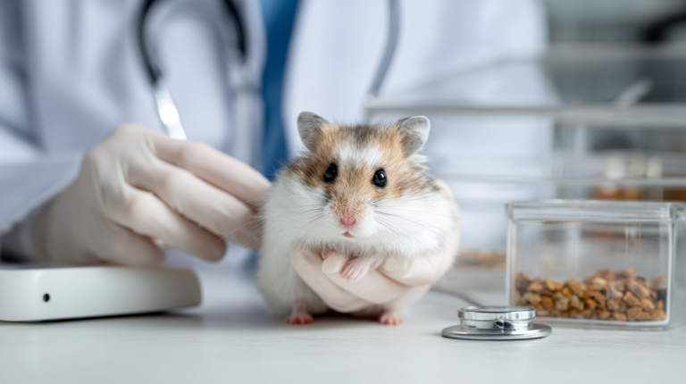
Diagnostic Approaches and Professional Assessment
When you suspect a respiratory infection in your rodent, professional veterinary diagnosis is essential. The subtle nature of early symptoms and the rapid progression of these infections make accurate diagnosis crucial for successful treatment.
Clinical Examination
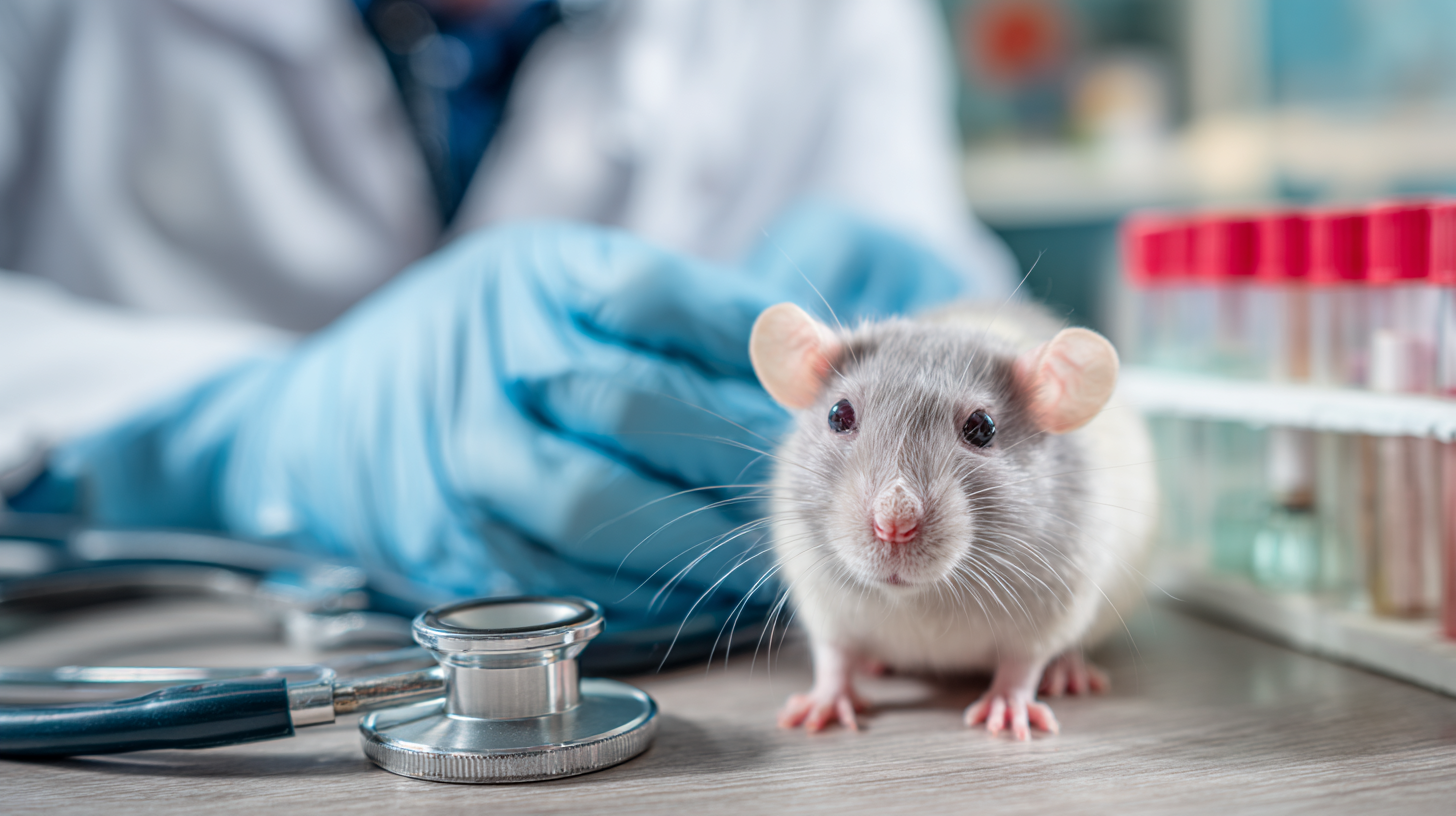
A thorough physical examination by an experienced exotic veterinarian forms the foundation of respiratory infection diagnosis. The veterinarian will assess breathing patterns, listen to lung sounds with a specialized stethoscope, and examine the nasal passages and throat.
Body weight measurement is particularly important, as respiratory infections often cause rapid weight loss due to increased energy expenditure and decreased appetite. The veterinarian will also evaluate body condition, hydration status, and overall demeanor.
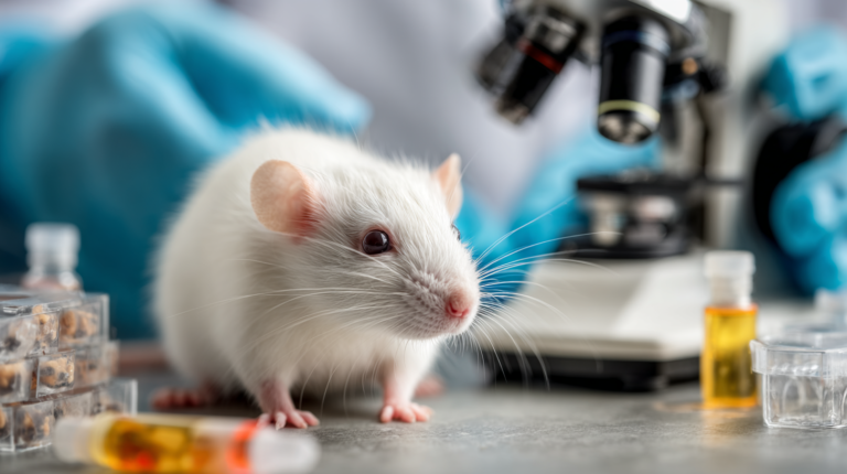
Advanced Diagnostic Tools
Radiography (X-rays) provides valuable information about lung health and can reveal pneumonia, fluid accumulation, or other structural abnormalities. Modern digital radiography allows for high-resolution images that can detect subtle changes in lung tissue.
Cytology and bacterial culture of nasal discharge or respiratory secretions can identify specific pathogens and determine antibiotic sensitivity. This information is crucial for selecting the most effective treatment and avoiding antibiotic resistance.
Blood work may reveal signs of infection, inflammation, or organ dysfunction. Complete blood counts can show elevated white blood cell counts indicative of infection, while chemistry panels can assess overall health status.
Differential Diagnosis
Respiratory infections must be differentiated from other conditions that can cause similar symptoms. Heart disease, particularly in older animals, can cause breathing difficulties that mimic respiratory infection. Allergic reactions to environmental factors can also cause respiratory symptoms.
Dental disease, particularly in guinea pigs and rabbits, can cause facial swelling and breathing difficulties that may be confused with respiratory infection. Tumors, though less common in young animals, can also cause respiratory symptoms.
Treatment Approaches and Management Strategies
| Treatment Phase | Timeline | Diagnostic/Treatment Actions | Expected Outcomes | Next Steps |
|---|---|---|---|---|
Initial Assessment |
0-2 hours |
Physical examination and breathing assessment
Temperature and weight measurement
Basic blood work if stable enough
Chest X-rays if indicated
|
Diagnosis confirmed Severity assessment completed |
Begin immediate treatment |
Emergency Stabilization |
2-6 hours |
Oxygen therapy if severe respiratory distress
Fluid therapy for dehydration
Start broad-spectrum antibiotics
Supportive care (warmth, nutrition)
|
Breathing stabilized Patient more comfortable |
Continue monitoring and treatment |
Acute Treatment |
Day 1-3 |
Continue antibiotic therapy
Nebulization therapy if needed
Pain management if required
Daily monitoring of vital signs
Common antibiotics: Enrofloxacin, Trimethoprim-sulfa
|
Symptoms improving Appetite returning |
Adjust treatment based on response |
Response Evaluation |
Day 3-5 |
Assess treatment response
Culture and sensitivity if not improving
Modify antibiotics if needed
Continue supportive care
|
Significant improvement or Requires treatment adjustment | Continue current protocol or modify |
Recovery Phase |
Day 5-10 |
Complete antibiotic course (7-14 days total)
Gradual reduction of supportive care
Monitor for secondary complications
Environmental management review
|
Normal breathing restored Full activity level returning |
Plan follow-up care |
Follow-up Monitoring |
Week 2-4 |
Post-treatment examination
Ensure complete resolution
Address any lingering symptoms
Implement prevention strategies
|
Full recovery confirmed No recurrence of symptoms |
Regular health monitoring |
Prevention Focus |
Ongoing |
Environmental optimization
Stress reduction measures
Quarantine protocols for new animals
Regular health checks
|
Reduced reinfection risk Improved overall health |
Maintain preventive measures |
Successful treatment of respiratory infections in rodents requires a multi-faceted approach combining antimicrobial therapy, supportive care, and environmental management. The specific treatment plan depends on the severity of infection, identified pathogens, and the individual animal’s condition.
Antimicrobial Therapy
Antibiotic selection for rodent respiratory infections requires careful consideration of pathogen susceptibility, drug safety, and penetration into respiratory tissues. Enrofloxacin, a fluoroquinolone antibiotic, is commonly used for its broad-spectrum activity and excellent tissue penetration.
Trimethoprim-sulfamethoxazole combinations are often effective against common respiratory pathogens and are generally well-tolerated by rodents. For severe infections or when Mycoplasma is suspected, doxycycline may be preferred due to its specific activity against these organisms.
Treatment duration typically ranges from 7-14 days, but may be extended for chronic or severe infections. It’s crucial to complete the full course of antibiotics even if symptoms improve, as premature discontinuation can lead to treatment failure and antibiotic resistance.
Supportive Care
Supportive care often determines the outcome of respiratory infection treatment. Oxygen therapy may be necessary for animals with severe breathing difficulties. This can be provided through oxygen cages, nasal cannulae, or face masks, depending on the animal’s tolerance and condition.
Fluid therapy addresses dehydration that commonly accompanies respiratory infections. Subcutaneous or intravenous fluids may be administered, with careful monitoring to avoid fluid overload that could worsen breathing difficulties.
Nutritional support is critical, as sick animals often have reduced appetites. Hand-feeding with nutritious, easily digestible foods or commercial critical care formulas may be necessary. Vitamin C supplementation is particularly important for guinea pigs.
Environmental Modifications
Improving the cage environment is essential for recovery and prevention of reinfection. Enhanced ventilation reduces pathogen load and removes irritating gases. However, avoid creating drafts that could further stress the respiratory system.
Humidity control can help reduce respiratory irritation. Slightly increased humidity (50-60%) can help keep respiratory secretions moist and easier to clear, but excessive humidity can promote bacterial growth.
Temperature regulation ensures optimal comfort and reduces metabolic stress. Maintaining temperatures in the upper range of normal for the species can help support immune function and reduce energy expenditure.
Prevention Strategies for Long-term Health
Preventing respiratory infections is far more effective than treating them. A comprehensive prevention program addresses environmental factors, nutrition, stress reduction, and early detection of potential problems.
Environmental Management
Cage design and ventilation form the foundation of respiratory health. Cages should provide adequate space, proper ventilation without drafts, and easy cleaning access. Wire-bottom cages should be avoided as they can cause foot injuries and don’t provide adequate protection from drafts.
Bedding selection significantly impacts respiratory health. Paper-based bedding, hemp bedding, or aspen shavings are preferred over cedar or pine. Bedding should be dust-free and changed regularly to prevent ammonia buildup.
Air quality monitoring includes regular assessment of ammonia levels, dust accumulation, and overall ventilation effectiveness. Simple test strips can measure ammonia levels, with anything above 25 ppm requiring immediate attention.
Nutritional Optimization
Species-appropriate diets provide the nutritional foundation for strong immune function. Commercial rodent foods should be fresh, stored properly, and supplemented with fresh vegetables and fruits as appropriate for the species.
Vitamin C supplementation is crucial for guinea pigs, with requirements of 30-100mg daily depending on age and reproductive status. Fresh vegetables high in vitamin C, such as bell peppers and leafy greens, should be provided daily.
Probiotic supplementation may help maintain healthy gut flora and support immune function, particularly during antibiotic treatment or times of stress.
Stress Reduction
Appropriate housing includes adequate space, hiding places, and environmental enrichment. Social species should be housed in compatible groups, while solitary species should have sufficient space and stimulation.
Routine consistency helps reduce stress by providing predictable care schedules. Feeding, cleaning, and handling should occur at consistent times to minimize disruption to the animal’s natural rhythms.
Gradual changes to diet, housing, or environment help prevent stress-induced immunosuppression. Any necessary changes should be implemented slowly over several days to weeks.
Early Detection Programs
Regular health checks should include weekly weight measurements, daily observation of breathing patterns, and assessment of activity levels. Maintaining a simple log can help identify subtle changes that might indicate developing problems.
Quarantine protocols for new animals help prevent introduction of respiratory pathogens to established colonies. New animals should be housed separately for 2-4 weeks and observed for signs of illness before introduction to other animals.
Emergency Response and When to Seek Help
Recognizing when a respiratory infection requires emergency veterinary care can save your pet’s life. Certain signs indicate imminent danger and require immediate professional intervention.
Emergency Warning Signs
Cyanosis (blue-tinged lips, gums, or tongue) indicates severe oxygen deprivation and requires immediate emergency care. This sign suggests that the respiratory system is failing to provide adequate oxygen to the body’s tissues.
Collapse or extreme lethargy where the animal is unresponsive or barely responsive indicates severe systemic illness. Animals in this condition require immediate intensive care and have a guarded prognosis.
Severe breathing distress with open-mouth breathing, extreme effort, or inability to rest comfortably requires emergency evaluation. These signs suggest impending respiratory failure.
Finding Qualified Veterinary Care
Exotic animal veterinarians have specialized training in rodent medicine and are best equipped to handle respiratory infections. Not all veterinarians have experience with small mammals, so it’s important to identify qualified practitioners before emergencies arise.
Emergency clinics that see exotic animals should be identified and their contact information readily available. Some areas have specialized exotic animal emergency services that operate after hours.
Telemedicine consultations with exotic veterinarians can provide guidance when immediate in-person care isn’t available, though they cannot replace hands-on examination and treatment.
Preparing for Veterinary Visits
Documentation of symptoms, including videos of breathing patterns and detailed notes about when symptoms began, helps veterinarians make accurate diagnoses quickly.
Transport considerations for sick animals include maintaining appropriate temperature, minimizing stress, and ensuring adequate ventilation during transport. Sick animals should be kept warm and quiet during transport.
Treatment history including any medications given, changes in diet or environment, and previous health issues provides crucial information for veterinary assessment.
The infographic would display a central rodent figure with arrows pointing to each of the seven warning signs, accompanied by visual representations of normal versus abnormal breathing patterns. A side panel would show the progression from early warning signs to emergency symptoms, with clear action steps for pet owners at each stage.
Frequently Asked Questions
Expert answers about respiratory infections in rodents
Respiratory infections in rodents can progress extremely rapidly, sometimes within 24-48 hours from first symptoms to life-threatening conditions. This rapid progression is due to their small size, high metabolic rate, and delicate respiratory anatomy. Early intervention is crucial for successful treatment outcomes.
Yes, many respiratory pathogens can spread between different rodent species. Bacteria like Bordetella and Pasteurella are particularly capable of cross-species transmission. This is why quarantine protocols are essential when introducing new animals, and why sick animals should be isolated immediately.
While environmental improvements like increased humidity and better ventilation can provide some comfort, respiratory infections in rodents require professional veterinary treatment with appropriate antibiotics. Home remedies alone are insufficient and can delay critical treatment, potentially resulting in death.
Normal breathing in rodents should be quiet, regular, and through the nose. Breathing rates vary by species but should be consistent during rest periods. Any audible breathing sounds, mouth breathing, or visible effort indicates abnormal breathing requiring veterinary evaluation.
Upper respiratory infections primarily affect the nose, sinuses, and throat, causing symptoms like nasal discharge and sneezing. Lower respiratory infections involve the lungs and cause more severe symptoms like pneumonia, with greater risk of life-threatening complications.
Stress doesn’t directly cause respiratory infections, but it significantly weakens the immune system, making rodents more susceptible to infectious agents. Chronic stress can turn normally harmless bacteria into pathogenic organisms, leading to serious illness.
For more expert pet care tips and product recommendations, visit BlithePet.com — your trusted source for pet wellness.
Conclusion
Respiratory infections in rodents represent one of the most serious health challenges facing small pet owners. The seven critical breathing signs outlined in this guide – rapid shallow breathing, open-mouth breathing, audible breathing sounds, abnormal positioning, nasal discharge, reduced activity, and vocal changes – serve as your early warning system for detecting these potentially fatal conditions.
Remember that rodents are masters at hiding illness, and by the time symptoms become obvious, the infection may already be severe. This makes your role as an observant pet owner absolutely crucial. Regular health monitoring, optimal environmental conditions, and immediate veterinary care when warning signs appear can mean the difference between life and death for your beloved pet.
The key to successful management lies in prevention through proper husbandry, early detection through careful observation, and prompt professional treatment when infection occurs. Never underestimate the severity of respiratory symptoms in rodents – what might seem like a minor cold in other animals can quickly become fatal in these small, delicate creatures.
By staying vigilant, maintaining optimal care conditions, and developing a relationship with a qualified exotic veterinarian before emergencies arise, you can give your rodent companion the best possible chance for a long, healthy life free from respiratory complications.
Have a similar experience with your pet? Share it in the comments below!